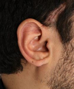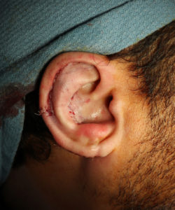
 This is a great example of what an ear looks like at the end of macrotia ear surgery.
There is obviously some degree of immediate surgical swelling and tissue distortion. But, overall, you can see more specifically how the upper portion of his ear has been reduced in size. The helical rim, in particular, is now much closer to the antihelix ridge. This is a direct reflection of the fact the scapha portion of the ear has now been reduced in size.
In this instance, Dr. Hilinski also reduced the size of his conchal bowl by taking out some of the cartilage and skin that were contributing to the ear looking larger than desired. The conchal bowl is the segment of the ear that forms a cup in the middle. Although this form of conchal bowl reduction was not extensive, every bit helps when it comes to making ears smaller in size!
As time goes on, the ear will begin to look much more normal in appearance. This includes resolution of the swelling and discoloration you see here.
